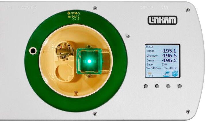
The CMS196 instrument, designed in association with Professor Bram Koster’s team from Leiden University Medical Centre (LUMC) in the Netherlands, is the outcome of four years of research and development.
相关阶段是出于使用荧光显微镜的工具的必要性而开发的,该工具可以帮助将感兴趣的结构定位在样品中,以进行高分辨率的冷冻电子显微镜成像。多年来.
A.J. Koster, Professor, Leiden University Medical Centre
CMS196V³CRYO-CLEM阶段:概述
Cryo-Clem仪器-CMS196V³-允许Clem的完整工作流程。它不仅可以通过液氮冷却来保持样品玻璃化,而且还建立了安全转移和处理冷冻样品并通过光学显微镜对其进行成像的能力。同时,它确保在任何给定时间中样品没有污染。
新模型的内置机动,编码阶段以及最新的链接软件有助于轻松,快速地创建网格的高分辨率图。这允许一项简单的坐标映射技术,以识别荧光显微镜和EM中的相同样品。
While the CMS196V³ instrument was particularly developed to resolve the concerns relating to vitrification of EM grids from the fluorescent microscope into the cryo TEM without contamination and devitrification through condensation, it also had to be improved optically to facilitate the use of high NA lenses.
Up to three grids are placed into a uniquely developed cassette, which, in turn, is shifted from a plunge freezer into a compact sealed container. This container is subsequently placed into a pre-cooled dry sample loading chamber fixed on an upright fluorescent microscope. Then, using unique manipulation tools, the cassette is easily placed onto the viewing bridge.
Features/Highlights of the CMS196V³
-
Automatic top-up liquid nitrogen is available to top up the chamber
-
独立的冷冻样品观察系统将标本玻璃体固定在-196°C的恒定温度下,并防止污染
-
Built-in motorized XY stage with position readout of better than 1 µm and optical encoders for high precision movements
-
Startup time takes only 10 minutes, no mechanical connection between Dewar and sample mount, excellent stability, good long-term stability and low drift in the nanometer range
-
A mounting option is available for a majority of research-grade, upright microscopes from all leading manufacturers
-
XY mapping is fully automated
-
支持自动电网(FEI),EM网格,贝西网格,膜载体(Leica HPM100),Croyocapcel的冷冻胶囊,Planchettes和定制的样品持有者;无需管理裸网格
-
A self-aligning magnetic sample cassette system is available for up to three sample grids
-
Can be linked to high pressure freezing and plunge-freezing sample preparation procedures
-
Sample cassette holder enables safe and contamination-free loading, transfer and storage of samples
-
内置冷凝器光学元件可实现阶段对比度和透射明亮场
-
Built-in sensors help monitor the temperature of samples
-
提供全天的个人操作,提供3-L自动填充LN2 Dewar作为选项
-
Fluorescence observation with up to 0.9 NA
-
自动加热“烘烤”周期消除了水分
-
带有按钮控制的LCD显示,通过USB接口进行完整的软件控制以及独立操纵杆操作
-
在世界各地的该领域已经证明了50多个系统,使Linkam Scientific成为Cryo-Clem领域的市场领导者
相关显微镜 - 高分辨率的EM网格的自动映射
CMS196M automated mapping of an EM grid at high resolution. Video Credit: Linkam Scientific Instruments
为什么为什么要冷冻?
While electron microscopy (EM) offers structural data at an excellent resolution, it can provide only a limited understanding of chemical and biological processes because of the limitations in sample preparation and staining procedures. By contrast, fluorescence microscopy is a highly sensitive technique that helps detect genetic, chemical and biological events and processes within living cells.
Now, Cryo-CLEM brings it all together: it is a novel, emerging method that integrates the individual benefits from both EM and fluorescence by imaging the location of the same specimen using both methods and superimposing the complementary data.
We are using the cryo-correlative stage to screen cells trapped in a thin layer of ice prior to imaging the samples at synchrotrons in Oxford, Barcelona and Berlin. The team at Linkam Scientific have helped to drive forward our research, quickly adapting and refining the technology to deliver an elegant, flexible, user-friendly stage.
Dr Lucy Collinson, Head of Electron Microscopy, The Francis Crick Institute
在冷冻条件下的玻璃样品成像
To make sure that a biological specimen is compatible with the vacuum conditions present in EM and that the structural detail is preserved, specimens are integrated into vitrified “glass-like” ice and these should be maintained below −140°C.
应防止与大气水分的任何接触,因为冰晶会迅速形成,从而污染样品。在冷冻条件下,保留荧光信号的结构细节,并大大减少光漂白。
Linkam Co-Ordinate Transform Tool

Image Credit: Linkam Scientific Instruments